Introduction of experimental autoimmune encephalomyelitis (EAE) model
Establishment of EAE mouse model
Experimental Animals:C57BL/6, 10-13 weeks old, female
Modeling reagent:MOG emulsion and PTX
Modeling method:Immunized with MOG emulsion and injected pertussis toxin intraperitoneally


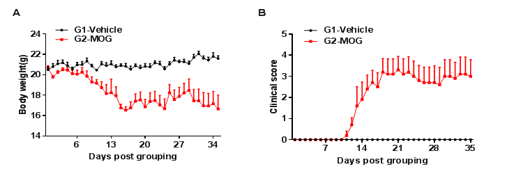
C57BL/6 mice (female, 8-week-old, n=5) immunized with MOG (myelin oligodendrocyte glycoprotein) emulsion, which were given PTX (pertussis toxin) on the day of immunization and the following day, respectively.(A) Body weight change of animals in each group. (B) Clinical scores of animals in each group. Compared with the untreated group (G1-Vehicle), the MOG-treated group (G2) had tail weakness, lameness, hind limb paralysis and other symptoms, resulting in an increased clinical score. This demonstrates that EAE was successfully induced in C57BL/6 mice. Data are shown as mean ± SEM.

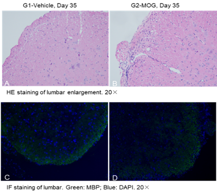
Local inflammatory responses in the central nervous system (CNS) of EAE model mice. Spinal cords were taken on day 35 after MOG and PTX immunization, and tissue sections were stained with H&E (A, B) and detected by immunofluorescence (IF) (C, D) (green: MBP; blue: DAPI). inflammatory cell infiltration (DAPI+ cells) was significantly increased and myelin protein was significantly reduced in the model group. This suggests that the EAE disease model was successfully induced in C57BL/6 mice.
Product list
|
Product name |
Product number |
|
B-hIL17A mice |
110053 |
|
B-hTNFA mice |
110002 |
References
1. Gaffen, S.L., Jain, R., Garg, A.V. & Cua, D.J. The IL-23-IL-17 immune axis: from mechanisms to therapeutic testing. Nat Rev Immunol 14, 585-600 (2014).
2. Iwakura, Y. & Ishigame, H. The IL-23/IL-17 axis in inflammation. J Clin Invest 116, 1218-1222 (2006).
3. Kuwabara, T., Ishikawa, F., Kondo, M. & Kakiuchi, T. The Role of IL-17 and Related Cytokines in Inflammatory Autoimmune Diseases. Mediators Inflamm 2017, 3908061 (2017).


