Nonalcoholic fatty liver disease (NAFLD) is a disease in which fat accumulates excessively in the liver. This fat accumulation is not caused by heavy alcohol consumption. Non-alcoholic steatohepatitis (NASH) is one type of NAFLD. In addition to liver fat accumulation, there are pathological changes of hepatitis, hepatocyte injury, fibrosis and liver scar formation, which may lead to cirrhosis or liver cancer. The mechanism of NASH progression remains unclear, and effective treatments are still lacking.
The clinical symptoms of NASH are quite complex and include obesity, insulin resistance, steatohepatitis, hepatocyte ballooning, and fibrosis according to the disease process. NASH models often used in preclinical experiments are genetic animal models, diet-induced animal models, and animal models constructed by combining genetics with diet-inductioned. However, it is difficult to mimic all the pathogenetic features of human diseases in a short period of time.
To understand the pathogenic mechanisms underlying the development and progression of NASH and to develop innovative therapies, we developed several mouse models of different stages of NASH pathogenesis.
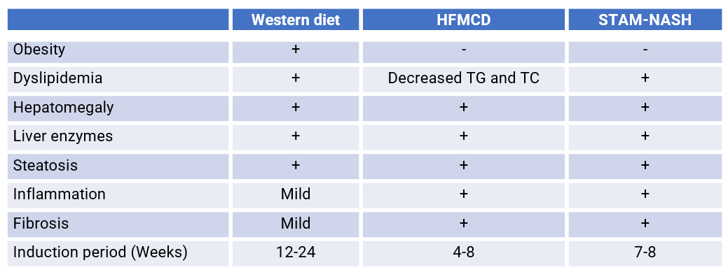
- Western diet (WD): Dietary components include high fructose and high cholesterol, and this diet-induced NASH model develops obesity, impaired glucose tolerance and hepatic steatosis.
- High-fat methionine-choline-deficient diet (HFMCD): The diet contains 60 kcal% fat, and is deficient in methionine and choline. , and this diet-induced NASH model shows increased liver injury, hepatic steatosis, and fibrosis accompanied by increased NAS scores.
- STAM-NASH: The Stelic Animal Model (STAM) mice are the first animal model of NASH-related liver carcinogenesis resembling disease development in humans.
The Western diet was a diet containing 21% fat, 50% carbohydrate, and 1.5% cholesterol, and this diet-induced NASH model developed obesity, impaired glucose tolerance, and hepatic steatosis.
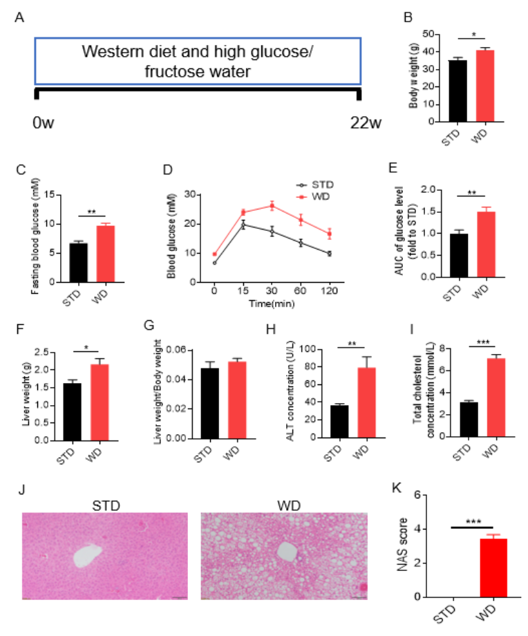
Western diet (WD) induced NASH model. Wild-type male C57BL/6J mice were randomly divided into 2 groups and given a standard diet (STD) ora Western diet (WD) diet consisting of high glucose and high fructose water, and various parameters were measured 22 weeks after feeding. (Fig. A) Schematic representation of Western diet (WD) -induced NASH mouse model; (B) body weight; (C) fasting glucose; (D) glucose tolerance test; (E) area under the curve of glucose tolerance test blood glucose level; (F) liver weight; (G) liver weight-to-body weight ratio; (H) serum ALT concentration; (I) serum total cholesterol; (J) H&E staining of liver sections; (K) NAFLD (non-alcoholic fatty liver disease) activity score (NAS). WT induced body weight gain,and increases in fasting blood glucose and glucose tolerance test blood glucose,increased liver weight, and an elevation in serum ALT and serum total cholesterol H&E staining indicates significant steatosis, and the NAS score was significantly increased. The above results indicate that WD can successfully establish a mouse model of NASH. Data are mean ± SEM, n = 8.
Efficacy Evaluation of the Anti-Glucagon Receptor (GCGR) Antibody Drug, Crotedumab, in a Mouse Model of NASH
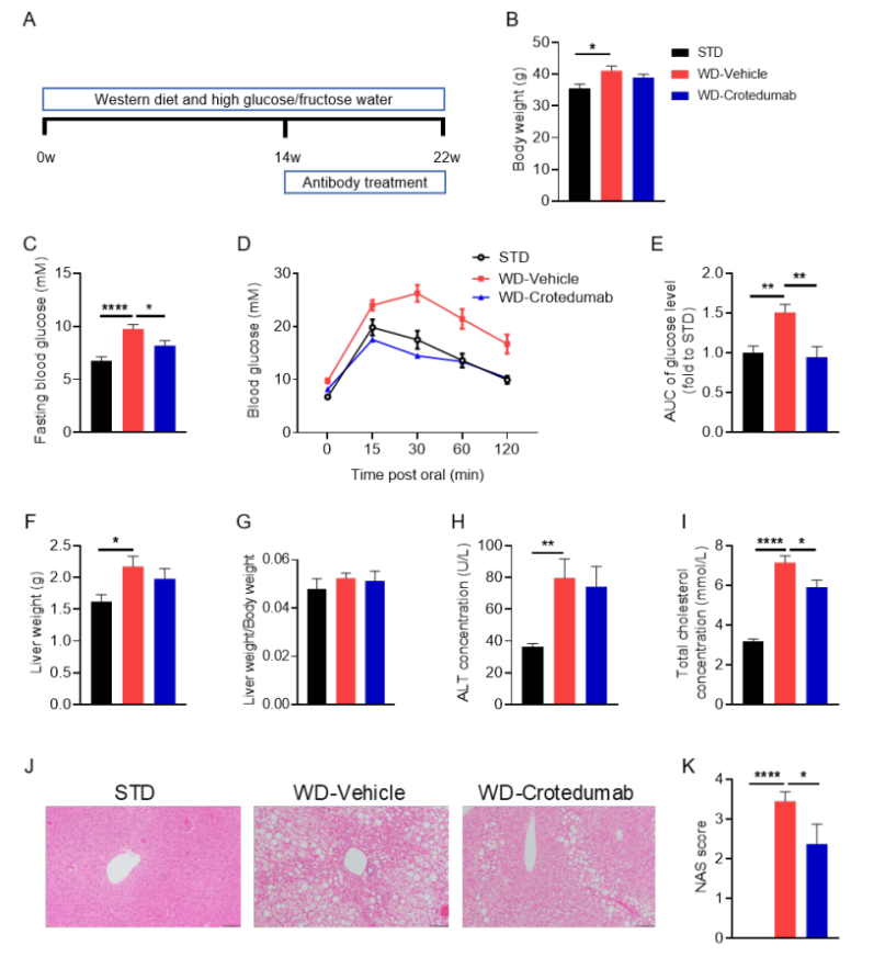
Crotedumab, a GCGR antibody, alleviated metabolic derangements in a mouse model of WD-induced NASH. ( A) Experimental schematic of NASH (non-alcoholic steatohepatitis) model establishment. Eight-week-old C57BL/6 male mice were fed a standard diet (STD) or a diet rich in cholesterol (Western diet; WD) containing 21% fat, 50% carbohydrate, and 1.5% cholesterol for 22 weeks. Blood glucose levels were analyzed and glucose tolerance tests were performed at week 13. Blood samples were collected at week 14 and analyzed for ALT and blood cholesterol, and blood samples were treated with rotedumab analog (synthesized in-house); (B) body weight; (C) fasting blood glucose; (D-E) glucose tolerance test and relative values of area under the curve; (F) liver weight; (G) ratio of liver weight to body weight; (H-I) serum ALT and total cholesterol concentrations; and (J-K) H&E staining and NAS scoring of liver tissue sections. The results indicate that drug administration significantly reduced fasting blood glucose and glucose tolerance. In addition,; liver weight and ALT were slightly decreased, serum total cholesterol concentration was decreased, and liver tissue steatosis and ballooning were relieved. The above results indicate that the application of glucagon receptor (GCGR) antibody can significantly reduce the symptoms of non-alcoholic steatohepatitis induced by Western diet. Data are mean ± SEM, n = 10.
References
1. Hernandez-Perez, E., Leon Garcia, P.E., Lopez-Diazguerrero, N.E., Rivera-Cabrera, F. & Del Angel Benitez, E. Liver steatosis and nonalcoholic steatohepatitis: from pathogenesis to therapy. Medwave 16, e6535 (2016).
2. Tsuchida, T., et al. A simple diet- and chemical-induced murine NASH model with rapid progression of steatohepatitis, fibrosis and liver cancer. J Hepatol 69, 385-395 (2018).
3. Farrell, G., et al. Mouse Models of Nonalcoholic Steatohepatitis: Toward Optimization of Their Relevance to Human Nonalcoholic Steatohepatitis. Hepatology 69, 2241-2257 (2019).
The HFMCD diet is a classic dietary model for NASH. Although the diet containsed 60 kcal% fat, it lacksed methionine and choline. Both methionine and choline can promote the delivery of fat from the liver through the blood in the form of phospholipids, improve the utilization of fatty acids in the liver, and prevent the abnormal accumulation of fat in the liver.
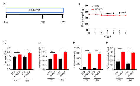
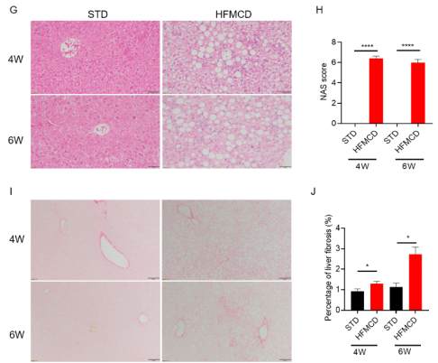
A model of NASH induced by a high-fat methionine-choline-deficient diet (HFMCD). Wild-type C57BL/6J mice were randomly divided into four groups and given a standard diet (STD) and a high-fat methionine-choline-deficient (HFMCD) diet and fed for 4 weeks and 6 weeks to detect various parameters. (Fig. A) HFMCD-induced NASH model protocol diagram; (B) mouse body weight; (C) liver weight; (D) ratio of liver weight to body weight; (E-F) serum ALT and AST concentrations; (G-H) HE staining and NAS score of liver tissue sections; (I-J) Sirius red staining of liver tissue sections and quantification statistics of the degree of liver fibrosis. The results showed that compared with the HFMCD-induced STD group, the liver weight was significantly increased; the serum ALT and AST concentrations were significantly increased. In addition, ; HE staining of liver tissue sections showed extensive hepatocyte steatosis, ballooning degeneration and intralobular inflammation; Sirius red staining of liver tissue showed significant liver fibrosis. The above results indicate that HFMCD induction can successfully establish a mouse model of NASH. Data are mean ± SEM, n = 5.
Efficacy Validation on NASH Mouse Models for celastrol
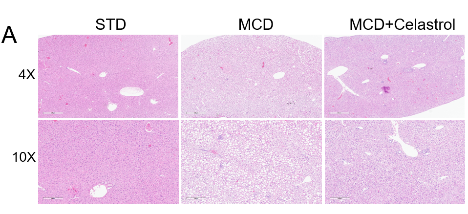
Analysis of H&E staining, index of liver function and NAFLD activity score the HFMCD-induced NASH mouse model. (A) Representative pictures of H&E staining showing reduced hepatic steatosis and inflammation after celastrol treatment. (B) ALT/AST change after MCD induction. Statistic data of NAFLD activity score (NAS). Values are expressed as mean ± SEM. **p<0.01, ***p<0.001.
References
1. Hernandez-Perez, E., Leon Garcia, P.E., Lopez-Diazguerrero, N.E., Rivera-Cabrera, F. & Del Angel Benitez, E. Liver steatosis and nonalcoholic steatohepatitis: from pathogenesis to therapy. Medwave 16, e6535 (2016).
2. Tsuchida, T., et al. A simple diet- and chemical-induced murine NASH model with rapid progression of steatohepatitis, fibrosis and liver cancer. J Hepatol 69, 385-395 (2018).
3. Farrell, G., et al. Mouse Models of Nonalcoholic Steatohepatitis: Toward Optimization of Their Relevance to Human Nonalcoholic Steatohepatitis. Hepatology 69, 2241-2257 (2019).
The Stelic Animal Model (STAM) mice are the first animal model of NASH-related liver carcinogenesis resembling disease development in humans1. It was found HCC developed in STAM mice is equivalent to stages B or C of the BCLC staging system in humans2. From a clinical perspective, animal experiments that mimic the development of human HCC will facilitate the development of new therapies directly related to human liver cancer, thus providing more successful opportunities for the prevention and treatment of HCC.
Establishment of STZ and HFD induced STAM-NASH Model
Exepriment Animal: C57BL/6, 0-24h, Male

In the STAM model, neonatal male mice were subcutaneously injected with 200μg streptozotocin (STZ) 2 days after birth and fed with high fat diet consisting of 60% of calories from fat starting at 4 weeks of age for 4 weeks.

Analysis of characteristics in STAM model of NASH. Neonatal male mice were injected with STZ on day 2 and fed a HFD diet from 4 weeks old. Body weight and blood glucose change after induction.
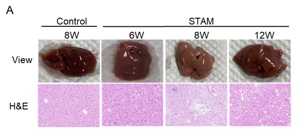
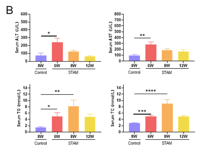
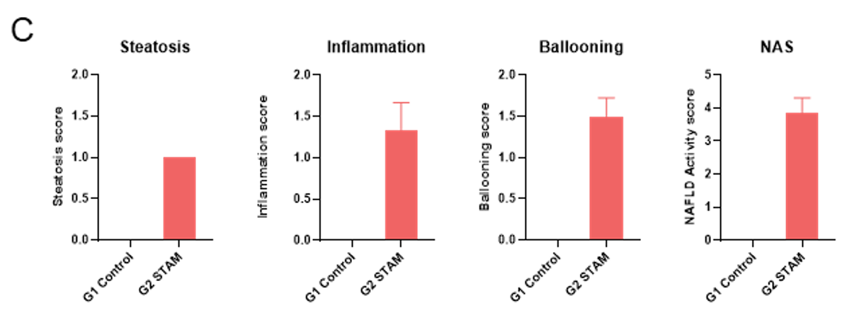
Analysis of characteristics in STAM model of NASH. (A) Representative pictures of H&E staining after 6, 8 and 12 weeks induction. (B) Blood biochemistry results showing the characteristics of STAM model. (C) NAFLD activity score. Values are expressed as mean ± SEM. *p<0.05, **p<0.01, ***p<0.001, ****p<0.0001.
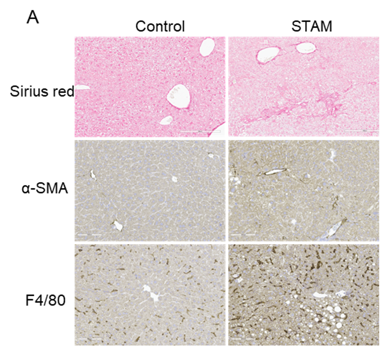
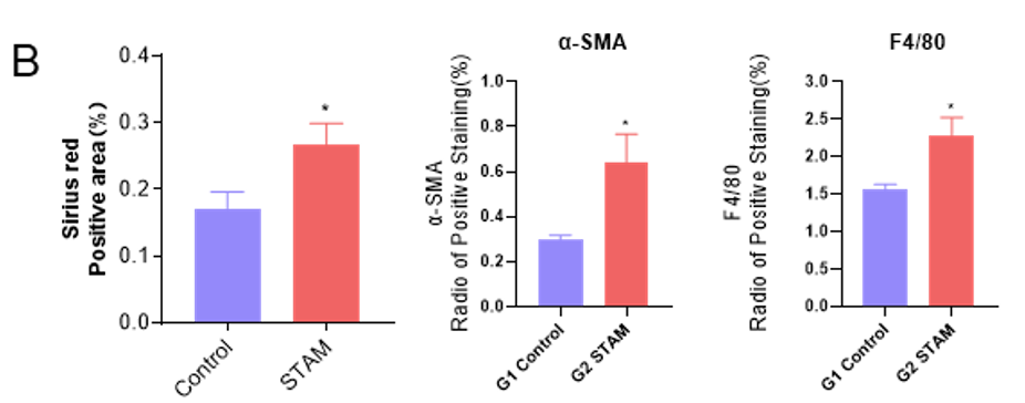
Analysis of fibrosis and inflammation in STAM model of NASH. (A) Representative pictures of Sirius red and IHC staining after 9 weeks induction. (B) Quantitative data of Sirius red and IHC staining. Values are expressed as mean ± SEM. *p<0.05.
Summary of diet and chemical induced NASH model

References
1. Tsuchida, T., et al., A simple diet- and chemical-induced murine NASH model with rapid progression of steatohepatitis, fibrosis and liver cancer. J Hepatol, 2018. 69(2): p. 385-395.
2 .Hansen, H.H., et al., Mouse models of nonalcoholic steatohepatitis in preclinical drug development. Drug Discov Today, 2017. 22(11): p. 1707-1718.
3. Ibrahim, S.H., et al., Animal Models of Nonalcoholic Steatohepatitis: Eat, Delete, and Inflame. Dig Dis Sci, 2016. 61(5): p. 1325-36.
4. Scholten, D., et al., The carbon tetrachloride model in mice. Lab Anim, 2015. 49(1 Suppl): p. 4-11.
5. Fujii M, et al. A murine model for non-alcoholic steatohepatitis showing evidence of association between diabetes and hepatocellular carcinoma. Med Mol Morphol. 2013;46:141–52.
6. Takakura K,. et al. Characterization of non-alcoholic steatohepatitis-derived hepatocellular carcinoma as a human stratification model in mice. Anticancer Res. 2014;34:4849–55.


